Metzler Veterinary Hospital

Orthopedic Surgery
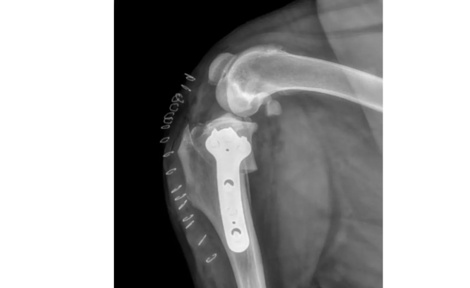
Cruciate ligament rupture (ACL/CCL tear)
One of the most frequent orthopedic conditions Dr. Dorian treats is the rupture of the cranial cruciate ligament (CCL/ACL). The most common surgery performed for this problem is a TPLO (tibial plateau leveling osteotomy). A special saw is used to cut the tibia (shin bone), this piece is rotated slightly and secured with a plate and screws. This procedure changes the physics of the knee, which allows your pet to walk much more comfortably even though the ligament is no longer present.
Another procedure can be performed for a CCL rupture, the Lateral Suture. This procedure is somewhat less invasive than the TPLO. However, it is usually reserved for smaller breed dogs (such as chihuahuas, yorkies, etc.), as medium/large/giant breed dogs tend to recover much better in the long run with a TPLO.
Medial Patellar Luxation (MPL)
Patellar luxation refers to a dislocated knee cap and is frequently seen in small-breed dogs; however, large-breed dogs (especially pit bulls) can suffer from this problem. This condition is congenital and developmental (your dog was born with this problem and it slowly got worse over time as he/she grew).
Correction of the problem can be quite complex and may involve multiple parts (deepening of the patellar groove, moving of the attachment point of the patellar ligament, soft tissue tightening/release, and sometimes correction of femoral curvature). This will be discussed with you at your pet’s consult.
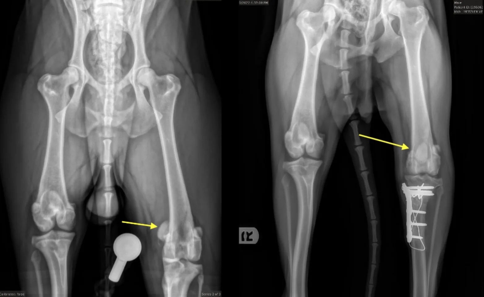
The dog in the image above had a combined CCL tear AND a medially luxating patella. When he came to us he was incredibly painful and would not even use his left leg. Dr. Dorian performed a combined TPLO and MPL procedure to stabilize this dog's knee. You can see the patella in an abnormal location (the yellow arrow) in the x-ray on the left.
In the x-ray on the right (after 10 weeks of healing) you can see the patella is now in a very normal location and this patient is doing incredibly well. You may have also noticed one femur is shorter than the other with a slight anatomic abnormality at one end, but this hasn’t caused our patient any difficulty. We are happy this pup has made an excellent recovery.
Fracture Repairs
A fracture is a break in a bone often caused by trauma, though sometimes other diseases/tumors can cause bones to break. If your pet has a fractured bone it is important that it is treated properly as improper treatment can lead to poor healing or chronic pain. There are various ways to treat a fracture, which are dependent on the type of fracture, bone, and patient. The treatment may range from splinting to plates and screws applied to the bone itself.
Dr. Dorian performs other orthopedic procedures as well such as FHOs (femoral head and neck ostectomy), hip luxation (dislocation) correction, arthrodesis (joint fusion), and surgery of other joints (although this is dependent on the type of issue your pet has). If you have any questions, please don’t hesitate to give us a call.
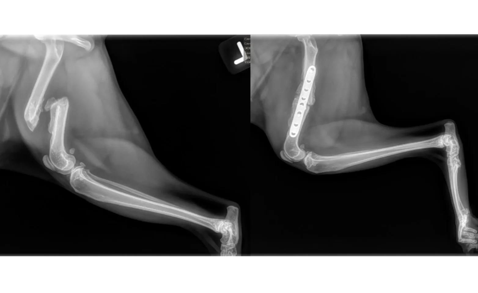
In the images above we see before and after pictures of a kitty's femur. He went missing for a month and came home with and fractured femur! The femur had actually begun the healing process; however, the bone was not healing in the proper location (the ends are very overlapped as you see here), and the kitty could not walk normally or without pain. A large amount of soft and hard callous wrapped around the bone and had to be broken down to re-align the fragments to allow placement of the plate. Within just a few days, this patient was walking again. The sent set of radiographs occurred about 4 weeks later and we can see new bone formation at the fracture site. This kitty still has a few more weeks before we can give the “all clear,” but given how well things have gone, we think he will do very well and heal with no complications.
